
Sign up to save your podcasts
Or

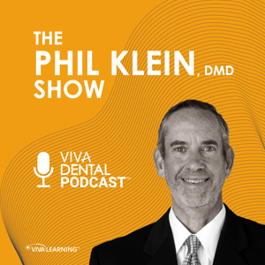
By Dr. Phil Klein | Dentist
The Dr. Phil Klein Dental Podcast is the #1 clinical dental podcast for evidence-based dentistry. Hosted by endodontist Dr. Phil Klein, DMD - trusted by 250,000+ dental professionals with 750+ epis
... more

4.7
3131 ratings


The podcast currently has 820 episodes available.
The most played episodes among Podcast App listeners.
What does it really take to preserve pulp vitality in a severely decayed posterior tooth — and how does your handpiece factor into that outcome? Dr. Anna Paholiouk is a restorative dentist practicing in Orange County, California, whose clinical philosophy is rooted in minimally invasive advanced adhesive dentistry based on biomimetic principles. She serves as Adjunct Faculty at the University of Southern California Herman Ostrow School of Dentistry — the same institution from which she graduated — contributing to the education and mentorship of the next generation of clinicians. Dr. Paholiouk holds the distinction of being the first and only female Fellow of the Academy of Biomimetic Dentistry, is a Fellow of the International Congress of Oral Implantologists, and has completed well over 1,000 hours of continuing education in pursuit of clinical excellence. In this episode, Dr. Paholiouk walks through her philosophy of circumscribing the pulp during preparation design, explaining how the goal of preserving nerve vitality dictates every clinical decision — from handpiece selection to isolation protocol. The conversation explores how electric handpieces have evolved to meet the demands of high-magnification, minimally invasive work, and why consistent handpiece maintenance is just as important as the handpiece itself. Dr. Paholiouk offers a remarkably systematic approach to clinical precision that reduces variables, prevents failures, and keeps patients out of the endodontic chair whenever biologically possible. Episode Highlights:Biomimetic preparation design centers on circumscribing the pulp chamber rather than removing structure for retention. The three-by-five rule — measuring three millimeters from the contact of an adjacent tooth and five millimeters from the occlusal surface — defines the anatomical zone where the pulp lives and guides how far preparations can safely extend while preserving nerve vitality.Electric handpieces offer a distinct clinical advantage over air-driven units because RPM can be controlled with a single bur throughout the entire preparation sequence. This allows the clinician to cut at maximum speed, then slow down to a low RPM on a dry field to finalize margins with the same fine diamond, reducing bur changes, improving efficiency, and giving greater tactile control during conservative preps.Consistent handpiece maintenance is critical to long-term performance, and automation is the most reliable way to eliminate human variability across staff members. Using an automated cleaning and lubrication system after every single procedure — rather than on a scheduled interval — ensures correct oil volume, proper internal gear rotation, and complete purging without relying on individual staff judgment, which has allowed one practice to go over five years without a single handpiece repair.Rubber dam isolation is non-negotiable in adhesive biomimetic dentistry because adhesion is chemistry, and chemistry requires a controlled environment. Placing a wingless clamp on the most posterior tooth and isolating the full quadrant eliminates saliva contamination, improves visibility with a contrasting dam color, protects the airway during bur use, and removes soft tissue interference — all of which directly support margin integrity and restoration longevity.Selective caries removal using a caries detection dye requires multiple reapplications and a clear understanding of what is being achieved. The goal is not to remove all stained dentin, but to establish a peripheral seal zone of approximately two to three millimeters of clean enamel, superficial dentin, and the dentinoenamel junction — leaving affected but not infected dentin in place when it is safely distant from the pulp to support remineralization and minimize iatrogenic pulpal exposure.Perfect for: General dentists looking to evolve their restorative philosophy toward more conservative, adhesive-based care; restorative specialists, dental residents, and dental students interested in biomimetic principles; and any clinician who wants to reduce handpiece-related failures through better maintenance protocols. If you have ever been told a tooth needs a root canal and a crown before anyone checked whether the pulp was actually vital, this episode will change the way you approach your next posterior restoration.

Are you leaving serious revenue on the table by treating your hygiene department as a break-even cost center? What if your hygienists could become the most powerful case acceptance drivers in your entire practice? Katrina Klein is a registered dental hygienist with 17 years of clinical experience, national speaker, author, competitive bodybuilder, Certified Personal Trainer, Certified Ergonomic Assessment Specialist, and Functional Range Conditioning Mobility Specialist. She is the founder of ErgoFitLife, an organization dedicated to making ergonomics and fitness a lifestyle for dental professionals to prevent, reduce, or eliminate occupational pain. With nearly 30 years in dentistry and a reputation as a leading voice in dental hygiene advocacy, Katrina brings a uniquely evidence-informed, practice-centered perspective to expanding the hygiene role. In this episode, Dr. Phil Klein and Katrina Klein challenge the widely held belief that the dental hygiene department is a loss leader, arguing instead that it can and should be a significant revenue-generating hub within any practice. The conversation covers how hygienists can move beyond scaling, root planing, and prophy to incorporate soft tissue laser therapy, adjunctive periodontal therapies, oral probiotic recommendations, airway assessment, implant maintenance, and orthodontic case co-diagnosis. They also address the current HR crisis in dental hygiene, the growing threat of DSO models utilizing minimally trained personnel for hygiene-adjacent procedures, and the critical importance of philosophical alignment between hygienists and practice owners. This discussion is essential for any dental team serious about elevating patient care and practice profitability simultaneously. Episode Highlights:A hygiene department focused solely on prophylaxis and scaling and root planing is unlikely to cover its own overhead at current wage rates. By incorporating comprehensive assessments, adjunctive periodontal therapies, airway screening, oral cancer screening, and co-diagnosis of restorative needs, hygienists can transform their operatory into a consistent revenue generator beyond insurance reimbursement ceilings.A real-world case was presented in which a non-compliant periodontal patient was introduced to an adjunctive peroxide-based tray delivery system, resulting in reduced calculus burden, improved periodontal health, tooth whitening, and ultimately full patient buy-in for clear aligner therapy and soft tissue laser treatment at every maintenance visit — all within an insurance-based practice.Soft tissue laser therapy is identified as one of the highest-impact tools available to hygienists, yet lasers frequently sit unused in practices because hygienists lack hands-on training and confidence. Sending the hygiene team together to live, hands-on continuing education courses — rather than passive lecture-format CE — is recommended as the most effective strategy for clinical adoption and team-wide implementation.Peri-implantitis is described as a growing clinical priority, with hygienists now encountering implant patients at nearly every maintenance appointment in periodontal-focused practices. Updated protocols have replaced outdated instrumentation approaches, and ongoing continuing education is emphasized as essential for hygienists to confidently probe, assess, and educate implant patients on proper home care.A legislative trend is emerging in multiple states allowing dental assistants with as few as 120 hours of training to perform supragingival scaling under DSO models, creating a direct workforce and quality-of-care challenge for registered dental hygienists. Hygienists are encouraged to proactively demonstrate their expanded clinical value, advocate for their scope of practice, and educate patients on the meaningful difference between credentialed hygiene care and minimally trained scaling services.Perfect for: Dental hygienists at all career stages looking to expand their clinical role and earning potential, general dentists and practice owners evaluating hygiene department productivity, and dental residents or new graduates building their philosophy around comprehensive team-based care. If you have ever wondered whether your hygiene department is working as hard as it could for your patients and your practice, this episode will completely reframe how you think about it.
What if you could hand a patient their finished crown before they even leave the chair — and it cost you $22 to make? Chairside 3D printing is no longer a future concept; it is happening right now in high-volume practices, and it is reshaping how dentists think about same-day restorations. Dr. Michael Erdos is a general dentist and co-owner of Tawil Dental, a Brooklyn practice with nearly 50 years of history in cosmetic, restorative, and implant dentistry. A Columbia University College of Dental Medicine graduate and former Chief Resident at Mount Sinai Hospital, Dr. Erdos has built his clinical focus around the intersection of aesthetic dentistry, prosthodontics, and digital workflows. Over the past several years, he has worked directly with Shining3D to test, refine, and optimize in-office digital printing workflows at scale, with a particular emphasis on making AI-driven design and chairside printing practical for everyday clinical use. In this episode, Dr. Erdos walks through a complete chairside 3D printing workflow — from intraoral scan to cemented restoration — and explains exactly how a general dentist can deliver a crown, veneer, onlay, or pediatric crown in a single visit using a compact, countertop printer that weighs under five pounds and requires nothing more than a standard wall outlet. He compares chairside printing directly to milling, discusses the ceramic-infused resin materials now cleared for permanent restorations, and shares his honest assessment of where the aesthetics stand today versus lithium disilicate. This is one of the most clinically detailed and financially transparent conversations about chairside 3D printing available to the dental profession.Episode Highlights:The complete chairside workflow from scan to cemented restoration can be completed in approximately 30 minutes, with a significant portion delegable to a trained assistant. The process involves scanning the full arch and bite pre-operatively, scanning the preparation, uploading to a cloud-based platform, and using AI design software to generate a restoration proposal — typically within one to two minutes — before sending directly to the printer in a single click.Ceramic-infused resin materials now cleared for permanent restorations contain over 50% ceramic filler content, including ceramic compounds such as lithium disilicate and zirconia variants, which meaningfully increase strength and aesthetics compared to earlier-generation printable resins. While these materials do not yet rival the optical properties of pressed lithium disilicate or full-contour zirconia, post-cure staining, glazing, and nitrogen-atmosphere curing can significantly enhance final appearance and surface quality.The additive nature of 3D printing allows for margin and contour accuracy that can surpass subtractive milling in certain applications, particularly for thin restorations such as veneers, where burr diameter in milling limits the fineness of internal line angles and margins. For no-prep or minimal-prep veneer cases, printed restorations can be bonded over natural tooth structure with minimal or no enamel reduction, then refined chairside after cementation.From a financial standpoint, the per-restoration material cost using sealed single-use resin capsules is approximately $22, and a single capsule can accommodate multiple restorations of the same shade printed simultaneously — including up to six veneers or a three-unit bridge. At a practice fee of $750 per printed resin veneer, a four-veneer case generates approximately $3,000 in production at a material cost of $22, with the hardware investment in the printer recoupable within a single week of focused use.Chairside printing opens a clinically and financially accessible pathway for pediatric white crown restorations as a direct alternative to stainless steel crowns, allowing the printed crown to be fabricated during the same appointment while other restorative procedures are completed. The same workflow applies to budget-conscious adult patients currently treated with direct composite bonding, converting them to indirect printed resin veneers at a price point that improves case acceptance without competing directly with laboratory-fabricated porcelain veneer cases.Perfect for: General dentists exploring same-day digital workflows, prosthodontists integrating in-office printing, pediatric dentists seeking aesthetic alternatives to stainless steel crowns, and dental residents building their understanding of AI-assisted restorative design and chairside digital technology. If you have been waiting for 3D printing to become fast enough, affordable enough, and simple enough to work in a real practice — this episode will show you it already has.
Are you treating patients with dental implants the same way you treat natural teeth — and could that approach be putting those implants at risk? Dr. Jon Suzuki is a Professor of Microbiology and Immunology in the School of Medicine and Professor of Periodontology and Oral Implantology in the School of Dentistry at Temple University, where he also serves as Chairman and Program Director of the Department of Periodontology and Oral Implantology and Associate Dean for Graduate Education. A former Dean at the University of Pittsburgh and CEO of the University faculty practice plan, Dr. Suzuki holds a D.D.S. from Loyola University of Chicago, a Ph.D. in Microbiology from the Illinois Institute of Technology, an N.I.H. Fellowship in Immunology from the University of Washington, a Clinical Certificate in Periodontics from the University of Maryland, and an MBA from the Katz Graduate School of Business. He is a Diplomate of the American Board of Periodontology, a Diplomate and Board Examiner of the International Congress of Oral Implantology, a Boarded Specialist Microbiologist, a Fellow of the American and International College of Dentists, former Chairman of the FDA Dental Products Panel, and former Chairman of the ADA Council on Scientific Affairs, with over 150 published papers, chapters, and symposia and a textbook in Medical Technology. In this episode, Dr. Suzuki joins Dr. Phil Klein to deliver a comprehensive, evidence-based framework for managing peri-implant health in the general dental practice. The conversation spans the biological reasons implant disease progresses faster than periodontal disease around natural teeth, the emerging concern of titanium corrosion and its potential systemic implications, and the practical protocols hygienists should follow at every implant maintenance visit. With 5 million implants placed annually in the United States, this discussion is immediately relevant to nearly every dental professional seeing patients today. Episode Highlights:Because dental implants lack a periodontal ligament, they have no PDL-associated vascularity, diminished local immune response, and only an epithelial soft tissue collar rather than a full gingival fiber complex. These anatomical differences mean peri-implant disease can progress significantly faster than periodontitis around natural teeth, making early detection and intervention critical to implant survival.Probing depths of three to five millimeters are considered within normal limits around implants due to the nature of soft tissue adhesion, but pockets deepening to five to six millimeters, combined with erythema and bleeding on probing, represent clear red flags for peri-implant mucositis or early peri-implantitis. Hygienists should use a plastic or resin probe rather than a stainless steel probe during implant assessment to avoid scratching the titanium surface.Titanium corrosion from dental implants is an emerging area of clinical concern supported by parallel literature in orthopedics and obstetrics-gynecology. Corrosion risk increases once implant threads become exposed to the oral environment — accelerated by pH changes from plaque, dietary factors, and occlusal loading — and the potential for systemic distribution of corrosion byproducts warrants continued research and clinical vigilance.Approximately 80 to 85 percent of patients do not floss consistently, and implant patients are no exception. Power brushes, interproximal irrigating devices, and antimicrobial mouth rinses used selectively — particularly for two to three weeks following a surgical or maintenance appointment — represent practical, compliance-friendly strategies for reducing peri-implant biofilm in the home care setting.Smoking lowers sulcular oxygen tension, promotes pathogenic anaerobic bacteria, suppresses neutrophil response, reduces salivary flow, and diminishes secretory immunoglobulin A — all of which increase peri-implantitis risk. While smoking is not an absolute contraindication for implant placement, dental offices should offer a tiered cessation approach including community counseling, nicotine-containing lozenges and gums, nicotine patches, and — with significant caution given documented psychiatric adverse effects — prescription cessation medications.Perfect for: general dentists and restorative dentists managing implant patients, dental hygienists performing implant maintenance, periodontists, implant surgeons, and dental residents seeking evidence-based protocols for peri-implant disease prevention and early intervention. If you place, restore, or maintain implants — or if you have patients who do — this episode gives you a clinical framework you can apply at your very next appointment.
Are you maximizing your ability to save implants threatened by peri-implant disease? With millions of implants placed annually, peri-implant complications are becoming routine challenges in every practice. Dr. Robert Convissar brings over four decades of clinical experience and pioneering expertise in dental laser technology to this essential discussion. A New York City-based general practitioner, Dr. Convissar serves as Director of Laser Dentistry at New York Hospital Medical Center of Queens and has authored multiple textbooks including the acclaimed "Principles and Practices of Laser Dentistry," now in its third edition. An international lecturer across 30 countries, he has trained hundreds of dentists in laser applications and oral cancer detection techniques. This episode provides a comprehensive framework for incorporating laser therapy into peri-implant disease management. Dr. Convissar explains the critical distinction between peri-implant mucositis and peri-implantitis, emphasizing proper diagnosis of etiologic factors before any laser intervention. The discussion covers evidence-based protocols for post-scaling and root planing laser decontamination, optimal wavelength selection, and the essential role dental hygienists can play in laser-assisted therapy. Episode Highlights:Proper etiologic factor identification is mandatory before laser treatment, including evaluation of occlusal loading, crown contours, abutment integrity, and home care compliance. Without addressing these underlying causes, laser therapy provides minimal therapeutic benefit regardless of technique or wavelength selection.CO2 and erbium lasers operate at significantly lower tissue temperatures (100°C average tip temperature) compared to diode lasers (750-1500°C), making them safer choices for peri-implant therapy. This temperature differential reduces risk of thermal damage to implant surfaces and surrounding tissues while providing effective bacterial reduction.Laser therapy must function as an adjunct to conventional scaling and root planing rather than a replacement. The combination approach provides superior diseased tissue removal and logarithmic bacterial count reduction compared to mechanical debridement alone, leading to enhanced healing outcomes.Trained dental hygienists can effectively incorporate laser therapy into maintenance protocols when properly supervised and educated. This delegation expands treatment accessibility while creating additional revenue streams for practices already performing comprehensive peri-implant maintenance.Patient acceptance rates for laser-assisted peri-implant therapy are significantly higher than full-mouth periodontal laser treatment due to implant investment protection motivation. Single-implant laser treatment can serve as an introduction to expanded laser therapy applications throughout the mouth.Perfect for: General dentists, periodontists, oral surgeons, dental hygienists, and practice owners considering laser technology integration for enhanced peri-implant disease management and revenue diversification. Discover how laser technology can transform your approach to implant preservation and create sustainable practice growth opportunities.
The podcast currently has 820 episodes available.

39,081 Listeners

153,021 Listeners
13,482 Listeners

183 Listeners
390 Listeners
2,447 Listeners

71 Listeners
150 Listeners
20 Listeners

27,848 Listeners
265 Listeners

10,131 Listeners
19,066 Listeners

181 Listeners
6 Listeners